

Brachiopods (Phylum Brachiopoda) (Cambrian Present)
Benthic, sessile organisms which live in the sea with complex anatomy. Valves, with bilateral symmetry, are in carbonate and rarely in phosphate of calcium and the most important organs are in the posterior portion of the shell. They are the most abundant invertebrates in Paleozoic.Morphology
The most important their feature (Fif. 1) is surely an organ necessary for feeding, the lophophore. The typical brachiopod shell has one valve that is larger than the other (ventral), which has an opening (foramen) for a fleshy stalk called the pedicle, with which the brachiopod attaches itself to the substrate.The pedicle foramen may be enclosed on the anterior end by a single plate, called a deltidium, or by a pair of deltidial plates. The dorsal valve is known also as brachial, because the lophophore attaches to it (lophophore is also called "brachium", arm, since it has the same functions of human arm).
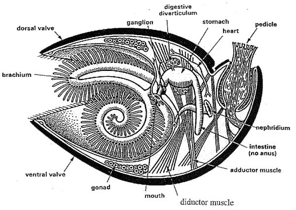
Fig.1: Brachiopod anatomy
Within the shell are two sets of muscles that open and close the shell (Fig.2) : adductors (run perpendicularly from the dorsal to the ventral valves and pull the two valves together, closing the shell) and diductors (insert to the middle of ventral valve and also on the cardinal process of the dorsal valve, so they pull the dorsal valve around its hinge line and cause it to open).
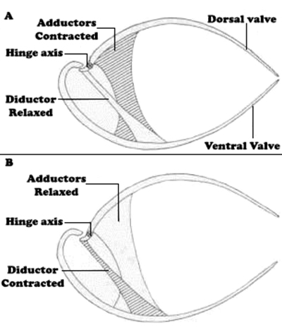
Fig.2: How brachiopods open and close valves
The external features of the brachiopod shell (Fig.3) may also be useful in classifying them. It can have a large flat or curved surface between the beak and the posterior margin of the other valve, which is known as interarea. The convex posterior portion of the shell is called umbo. The edge of shell along its line of closure (commissure) may be straight or have plication. Another interesting features are ornamentations ( mainly costae and growth lines).
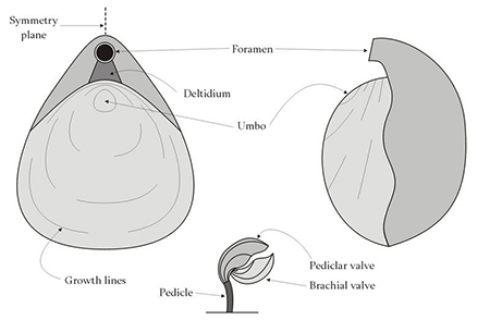
Fig.3: External features of the brachiopod shell
Brachiopods in thin section
Brachiopod microstructure is well visible in thin section. Usually it is fibrous, but sometimes it may be also prismatic. Observing brachiopods in thin section allows to define the conservation of shell and to describe possible modifications in the structure, which are very important for the classification.Uses in Paleontology
Their great abundance and diversity has made them very useful tools for paleoecology (sometimes its possible to study their biomineralization of the shell), biostratigraphy, evolutionary studies and paleogeography.page created by Dalila Grilli (Department of Earth Sciences, Milan, Italy)
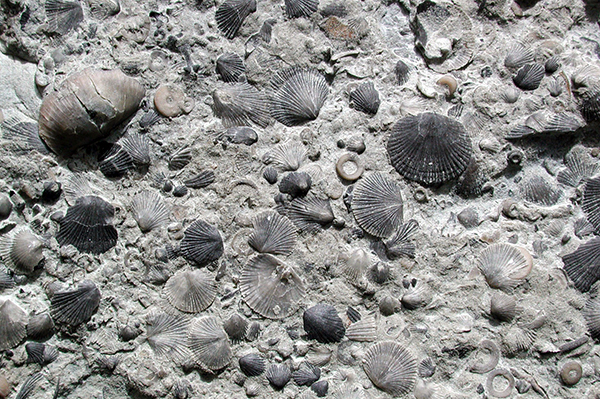
Brachiopods Fossils
Bibliography
• Prothero D.R., Bringing Fossils to Life, 2004 pp. 231 236
.jpg) Brachiopod shell with fibrous texture. PPL image, 10x (Field of view = 2mm) |
.jpg) Brachiopod shell with fibrous texture. XPL image, 10x (Field of view = 2mm) |
.jpg) Brachiopod shell with fibrous texture. XPL image, 10x (Field of view = 2mm) |
.jpg) Brachiopod shell with fibrous texture and external ornamentations. PPL image, 2x (Field of view = 7mm) |
.jpg) Brachiopod shell with fibrous texture and external ornamentations. XPL image, 2x (Field of view = 7mm) |
.jpg) Brachiopod shell with fibrous texture. XPL image, 2x (Field of view = 7mm) |
.jpg) Brachiopod shell with fibrous texture the dark lines inside the shell are "punctae". PPL image, 2x (Field of view = 7mm) |
.jpg) Brachiopod shell with fibrous texture. XPL image, 2x (Field of view = 7mm) |
.jpg) Brachiopod shell with fibrous texture. XPL image, 2x (Field of view = 7mm) |
.jpg) Brachiopod shell with fibrous texture. PPL image, 10x (Field of view = 2mm) |
.jpg) Brachiopod shell with fibrous texture. XPL image, 10x (Field of view = 2mm) |
.jpg) Brachiopod shell with fibrous texture. XPL image, 10x (Field of view = 2mm) |
.jpg) Brachiopod shell with fibrous texture. PPL image, 10x (Field of view = 2mm) |
.jpg) Brachiopod shell with fibrous texture. XPL image, 10x (Field of view = 2mm) |
.jpg) Brachiopod shell with fibrous texture. XPL image, 10x (Field of view = 2mm) |
.jpg) Brachiopod shell with fibrous texture. XPL image, 10x (Field of view = 2mm) |
.jpg) Brachiopod shell with fibrous texture. XPL image, 10x (Field of view = 2mm) |
.jpg) Brachiopod shell with fibrous texture. XPL image, 10x (Field of view = 2mm) |
.jpg) Brachiopods shell with fibrous texture and Fusuline (the black fossils). PPL image, 2x (Field of view = 7mm) |
.jpg) Brachiopod shell partially Phosphatized (the yellow-gray material). XPL image, 10x (Field of view = 2mm) |
.jpg) Brachiopod shell partially Phosphatized (the yellow material). PPL image, 10x (Field of view = 2mm) |
.jpg) Brachiopod shell partially Phosphatized (the yellow-gray material). XPL image, 10x (Field of view = 2mm) |
.jpg) Brachiopod shell partially Phosphatized (the yellow material). PPL image, 10x (Field of view = 2mm) |
.jpg) Brachiopod shell partially Phosphatized (the yellow-gray material). XPL image, 10x (Field of view = 2mm) |
.jpg) Brachiopod shell with Microboring structure (traces of predatory organisms). PPL image, 10x (Field of view = 2mm) |
.jpg) Brachiopod shell with Microboring structure (traces of predatory organisms). XPL image, 10x (Field of view = 2mm) |