

Veins
Veins are dilated fractures filled with oriented crystal fibers or non-oriented mineral deposits (typically quartz, calcite or carbonates). Veins occur in rocks of all types and metamorphic grades with thickness from less than a millimeter to several meters. Their morphology, petrology and chemistry are a valuable source of information in a range of geological disciplines. The association of many ore deposits (particularly gold) with veins makes them even more relevant to geology.The development of veins (Fig.1) is associated with the circulation of fluids in rocks, both for transport of material and for propagation and opening of the vein. The role of fluids in the formation and growth of veins is relatively complex. Fluid pressure may fluctuate in cycles of increasing pressure, leading to fracturing and vein opening, and subsequent falling pressure due to drainage during which mineral deposition commonly occurs. Some veins, however, show evidence of persistent high fluid pressure during mineral growth. Material deposited in a vein can be transported towards the dilatation site from outside in an open system along fracture networks, through the pore space, or along grain boundaries. This process is commonly referred to as advection and may cause changes in the chemical and isotope composition of a vein and its wall rock. Material deposited in veins can also be derived from the surrounding wall rock in a closed system, e.g. by dissolution and precipitation of quartz or calcite. In this case, material can be transported in a circulating fluid, or in a stationary fluid by diffusion.
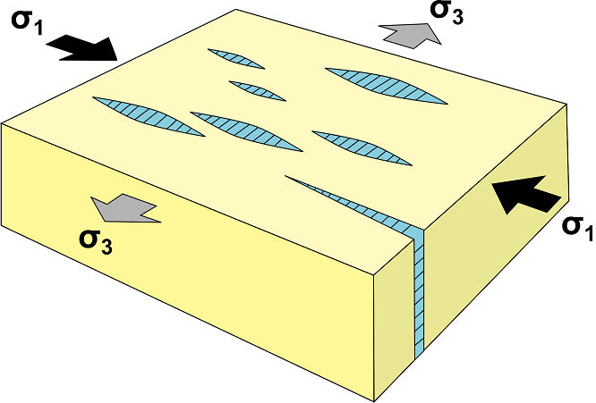
Fig.1: Vein orientation with respect to compression. Modified from Jean-Pierre Burg.
Crystals in veins
Many veins contain parallel oriented elongate crystals. This concerns naturally elongate minerals such as asbestos, actinolite or mica but more commonly minerals which do not normally have an elongate shape such as quartz, calcite, dolomite, feldspar, gypsum and. The elongate habit of minerals in such veins appears to be due to a special growth mechanism: Imagine a crack that forms in a crystalline aggregate of quartz or calcite. Immediately on opening, new crystalline material can be deposited on the existing crystals from solution in the fluid. If neighboring crystals grow at similar rates, the overgrowth obtains an elongate shape; crystals that grow slowly because they have an unsuitable crystallographic orientation will become thin and end while their neighbors make contact. A process of growth competition can lead to aggregates of few equidimensional grains, but if the growth competition is suppressed, many elongate grains will result.Veins classification
The terminology for veins that is currently in use, is mostly derived from Ramsay & Huber (1983) and Passchier & Trouw (1996). Terms for the description of veins can be grouped in three categories:- macroscopic morphology.
- microscopic morphology.
- growth morphology.
1) Macroscopic morphology: terms relating to macroscopic morphology, i.e. the shape of veins, are the well-defined. Broadly speaking, we can divide veins in two categories:
a) Pressure fringes: are veins that form on the two low pressure sides of hard objects, usually ore minerals, but also other objects, such as crinoid stem.
b) General veins: with shapes that are not primarily defined by a relatively hard object, but by fractures or other factors.
2) Microscopic morphology: the microscopic morphology relates to the texture or the shape and arrangement of crystals inside a vein. We can distinguish four primary categories: 1. Blocky crystals; 2. Elongate blocky crystals; 3. Fibrous crystals; 4. Stretched crystals.
Blocky texture: A blocky texture is a texture in which grains are roughly equidimensional and randomly oriented.
Elongate blocky texture: Crystals in an elongate blocky texture (Fisher & Brantley 1992) are typically moderately elongate and the long axes of crystals are aligned.
Fibrous texture: In a fibrous texture, the rod-shaped grains can achieve a much higher length/width ratio than in elongate blocky textures. As in an elongate blocky texture, the grainsâ long axes are aligned.
Stretched crystals: the primary distinction between the previous textures and stretched crystals is that in stretched crystals, additional growth took place inside the grains (on the surfaces of the half grains), with the space for new-growth provided by (micro-) fractures that cut through the grains.
3) Growth morphology: relates to where the site(s) of progressive growth are located in the vein, which determines the direction of growth of the vein forming crystals. Three types of growth direction of vein crystals relative to wall rock are identified:
• Syntaxial (inward) growth: Fibers grow from the wall in optical continuity with mineral grains of the same composition in the host rock (Fig.2). Fibrous growth can occur into the widening void from both sides at the same rate; this process can be referred to as bitaxial growth. Fibers extending from opposite walls meet at a medial suture where there is both a structural and optical discontinuity. The medial plane is the fracture plane where fiber separation is continuously sealed by new material added to both sides of the medial suture during successive cracking. Syntaxial veins are commonly asymmetric, i.e. with an off-centered median line, and in extreme cases grow from one side of the vein only. Such unitaxial veins lack a median line (Fig.3) .
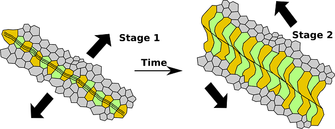
Fig.2: Development of Syntaxial vein. A change in the relative motion of the wall rocks (arrows) can cause curvature of the growing fibres in the veins. Modified from Passchier (2005).
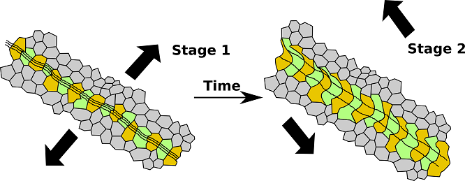
Fig.3: Development of Unitaxial vein. A change in the relative motion of the wall rocks (arrows) can cause curvature of the growing fibres in the veins. Modified from Passchier (2005).
• Antitaxial (outward) growth: Some veins are filled by growth of a mineral that is not the main constituent of the wall rock, e.g. a calcite vein in quartzite. In such cases, the growth usually occurs along the contact of elongate grains or fibres and the wall rock, i.e. on both sides of the material in the vein. A weak median line defined by small equidimensional grains or fragments of the wall rock is normally present in the center of the fibrous aggregate, indicating the initial nucleation site of the vein filling. This type of growth towards the wall rock is termed antitaxial growth, and the veins are called antitaxial veins (Fig.4). Material in antitaxial veins commonly consists of fibres rather than elongate crystals, and veins are commonly symmetric. Single fibres in antitaxial veins can be continuous over the median line, in contrast to elongate grains or fibres in syntaxial veins.
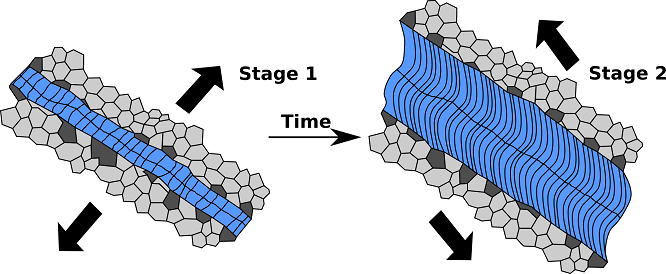
Fig.4: Development of Antitaxial vein. A change in the relative motion of the wall rocks (arrows) can cause curvature of the growing fibres in the veins. Modified from Passchier (2005).
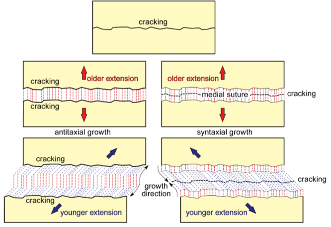
Fig.5: Mineral fibers parallel to the incremental opening direction of a Antitaxial and Syntaxial vein. From Jean-Pierre Burg.
• Ataxial growth: veins may form by repeated fracturing and growth at alternating different sites in the vein. Such non-localized, ataxial cracking and growth produces veins with jogged or stepped and rarely smooth elongate grains without a median line that are in continuity with fragments of single crystals on both sides of the vein. Such elongate grains are known as stretched crystals (Fig.6).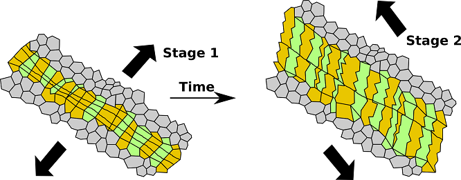
Fig.6: Development of Ataxial vein. A change in the relative motion of the wall rocks (arrows) can cause curvature of the growing fibres in the veins. Modified from Passchier (2005).
• Composite veins: are veins (Fig.7) in which an antitaxial vein segment is sandwiched between syntaxial vein rims. Such a vein has three median lines.
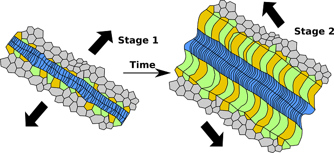
Fig.7: Development of Composite vein. A change in the relative motion of the wall rocks (arrows) can cause curvature of the growing fibres in the veins. Modified from Passchier (2005).
Bibliography
• Bons, P. D. (2000). The formation of veins and their microstructures. Journal of the Virtual Explorer, 2, 12.
• Bons, P. D., Elburg, M. A., & Gomez-Rivas, E. (2012). A review of the formation of tectonic veins and their microstructures. Journal of Structural Geology, 43, 33-62.
• Bucher, K., & Grapes, R. (2011). Petrogenesis of metamorphic rocks. Springer Science & Business Media.
• Fossen, H. (2016). Structural geology. Cambridge University Press.
• Howie, R. A., Zussman, J., & Deer, W. (1992). An introduction to the rock-forming minerals (p. 696). Longman.
• Passchier, Cees W., Trouw, Rudolph A. J: Microtectonics (2005).
• Philpotts, A., & Ague, J. (2009). Principles of igneous and metamorphic petrology. Cambridge University Press.
• Shelley, D. (1993). Igneous and metamorphic rocks under the microscope: classification, textures, microstructures and mineral preferred-orientations.
• Vernon, R. H. & Clarke, G. L. (2008): Principles of Metamorphic Petrology. Cambridge University Press.
• Vernon, R. H. (2018). A practical guide to rock microstructure. Cambridge university press.
.jpg) Big antitaxial quartz vein. Note the "median line" XPL image, 1x (Field of view = 9mm) |
.jpg) Big antitaxial quartz vein. Note the "median line" XPL image, 1x (Field of view = 9mm) |
.jpg) Big antitaxial quartz vein. Note the "median line" XPL image, 1x (Field of view = 9mm) |
.jpg) Big antitaxial quartz vein. Note the "median line" XPL image, 1x (Field of view = 9mm) |
.jpg) Big antitaxial quartz vein. Note the "median line" XPL image, 1x (Field of view = 9mm) |
.jpg) Big antitaxial quartz vein. Note the "median line" XPL image, 1x (Field of view = 9mm) |
.jpg) Big antitaxial quartz vein. Note the "median line" XPL image, 1x (Field of view = 9mm) |
.jpg) Big antitaxial quartz vein. Note the "median line" XPL image, 1x (Field of view = 9mm) |
.jpg) Big antitaxial quartz vein. Note the "median line" XPL image, 1x (Field of view = 9mm) |
.jpg) Particular of a big antitaxial quartz vein. Note the fluid inclusions within quartz crystals. PPL image, 2x (Field of view = 7mm) |
.jpg) Particular of a big antitaxial quartz vein. Note the fluid inclusions within quartz crystals. XPL image, 2x (Field of view = 7mm) |
.jpg) Particular of a big antitaxial quartz vein. Note the fluid inclusions within quartz crystals. XPL image, 2x (Field of view = 7mm) |
.jpg) Epidote vein in a Hornfels. Monte cristo island, Italy. PPL image, 2x (Field of view = 7mm) |
.jpg) Epidote vein in a Hornfels. Monte cristo island, Italy. XPL image, 2x (Field of view = 7mm) |
.jpg) Epidote vein in a Hornfels. Monte cristo island, Italy. PPL image, 2x (Field of view = 7mm) |
.jpg) Epidote vein in a Hornfels. Monte cristo island, Italy. PPL image, 2x (Field of view = 7mm) |
.jpg) Epidote veins in a Hornfels. Monte cristo island, Italy. XPL image, 2x (Field of view = 7mm) |
.jpg) Epidote vein in a Hornfels. Monte cristo island, Italy. XPL image, 2x (Field of view = 7mm) |
.jpg) Syntaxial quartz vein. XPL image, 10x (Field of view = 2mm) |
.jpg) Syntaxial quartz vein. XPL image, 10x (Field of view = 2mm) |
.jpg) Syntaxial quartz vein. XPL image, 10x (Field of view = 2mm) |
.jpg) Syntaxial quartz vein. XPL image, 10x (Field of view = 2mm) |
.jpg) Syntaxial quartz vein. XPL image, 10x (Field of view = 2mm) |
.jpg) Syntaxial quartz vein. XPL image, 10x (Field of view = 2mm) |
.jpg) Antitaxial quartz vein i a schist. XPL image, 10x (Field of view = 2mm) |
.jpg) Antitaxial quartz vein in a schist. XPL image, 10x (Field of view = 2mm) |
.jpg) Antitaxial quartz vein in a schist. XPL image, 10x (Field of view = 2mm) |
 Antitaxial quartz vein in a schist. XPL image, 10x (Field of view = 2mm) |
.jpg) Syntaxial quartz vein. XPL image, 10x (Field of view = 2mm) |
.jpg) Syntaxial quartz vein. XPL image, 10x (Field of view = 2mm) |
.jpg) Syntaxial quartz veins. XPL image, 10x (Field of view = 2mm) |
.jpg) Antitaxial quartz vein in a marble. XPL image, 10x (Field of view = 2mm) |
.jpg) Calcite vein in a marble. XPL image, 10x (Field of view = 2mm) |
.jpg) Calcite vein in a quartzite. XPL image, 10x (Field of view = 2mm) |
.jpg) Syntaxial quartz veins. From Pisa (Italy) XPL image, 10x (Field of view = 2mm) |
.jpg) Syntaxial quartz veins. From Pisa (Italy) XPL image, 10x (Field of view = 2mm) |
.jpg) Syntaxial quartz veins. From Pisa (Italy) XPL image, 10x (Field of view = 2mm) |
.jpg) Syntaxial quartz veins. From Pisa (Italy) XPL image, 10x (Field of view = 2mm) |
.jpg) Syntaxial quartz veins. From Pisa (Italy) XPL image, 10x (Field of view = 2mm) |
.jpg) Fibrous calcite crystals in a vein. Serpentinite from Monterosso Calabro, Calabria region, Italy. PPL image, 10x (Field of view = 2mm) |
.jpg) Fibrous calcite crystals in a vein. Serpentinite from Monterosso Calabro, Calabria region, Italy. XPL image, 10x (Field of view = 2mm) |
.jpg) Fibrous calcite crystals in a vein. Serpentinite from Monterosso Calabro, Calabria region, Italy. PPL image, 10x (Field of view = 2mm) |
.jpg) Fibrous calcite crystals in a vein. Serpentinite from Monterosso Calabro, Calabria region, Italy. XPL image, 10x (Field of view = 2mm) |
.jpg) Fibrous calcite crystals in a vein. Serpentinite from Monterosso Calabro, Calabria region, Italy. XPL image, 10x (Field of view = 2mm) |
.jpg) Fibrous calcite crystals in a vein. Serpentinite from Monterosso Calabro, Calabria region, Italy. XPL image, 20x (Field of view = 1mm) |
Fibrous calcite crystals in a vein. Serpentinite from Monterosso Calabro, Calabria region, Italy. XPL image, 20x (Field of view = 1mm) |
.jpg) Fibrous calcite crystals in a vein. Serpentinite from Monterosso Calabro, Calabria region, Italy. PPL image, 20x (Field of view = 1mm) |
.jpg) Fibrous calcite crystals in a vein. Serpentinite from Monterosso Calabro, Calabria region, Italy. XPL image, 20x (Field of view = 1mm) |