

Pseudotachylyte
Friction of rock against rock during an earthquake cause heating: frictional heating. When this happens at the base of the brittle zone, the rocks are already warm (few hundreds of degrees) and the additional frictional heating can lead to melting of the rock. An earthquake event is very short (seconds) and the molten rock quickly cools again to form a black glass, called Pseudotachylyte, on the fault plane or squirted into other cracks . Pseudotachylytes typically form at the brittle-ductile transition, where rocks are hot enough, but also still brittle enough. Pseudotachylyte is a cohesive glassy or very fine-grained fault-rock with a very distinct fabric . Its curious name derives from its resemblance to tachylyte, a mafic volcanic glass, while the material is obviously not of volcanic origin.Pseudotachylyte has a number of characteristic geometric features that usually allow its distinction from other brittle fault rock types. It is composed of a dark matrix material with minor inclusions of mineral or wall rock fragments. It usually occurs in a characteristic geometrical setting of a planar main fault vein or generation surface, up to a few mm thick and irregular injection veins which branch from the main fault vein into the wall rock. They never show transitional zones of decreasing brittle deformation intensity towards the wall rock as is usually the case for cataclasite or breccia.
Pseudotachylyte is thought to form by local melting of the rock along a brittle fault plane due to heat generated by rapid frictional sliding (102 to 1 ms1; Philpotts 1964). Pseudotachylyte occurs associated with events such as meteorite impact crater, crater collapse, caldera collapse and giant landslides on superficial superfaults in veins thicker than 1 cm.
Melting at temperatures between 7501 600 °C is thought to occur on the main fault vein of a pseudotachylyte. Some of the melt may intrude minor faults, which branch from the main fault vein into the wall rock, and form injection veins. The small volume of melt formed in this way cools rapidly to the temperature of the host rock. As a result, the melt quenches to a glass or very fine-grained, aphanitic material that occurs along fault planes and adjacent branching injection veins.
The matrix of pseudotachylyte is commonly black, dark brown, green or red and relatively homogeneous, but may contain a compositional layering of irregular thickness, which follows the contours of the vein. This layering is commonly of a different colour along the vein wall and in the interior, and is interpreted to result from selective melting of the wall rock. The layering may be folded and folds are interpreted to have formed by fluid flow in the melt.
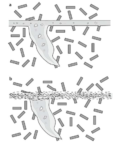
a Schematic drawing of a typical pseudotachylyte with main fault vein, injection vein, internal compositional banding and typical inclusions. The boundary with the wall rock is sharp. Mica grainsin the wall rock show corrosion along the contact with pseudotachylyte. b Pseudotachylyte in which the main fault vein has been reactivated as a mylonite zone. The mylonite can be recognised as a former pseudotachylyte by its fine-grained homogeneous nature and the presence of injection vein relicts. From Microtectonics (2005).
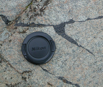
pseudotachylyte injection vein in the Balmuccia Peridotite, Ivrea-Verbano (Italy). From Francesca Meneghini.
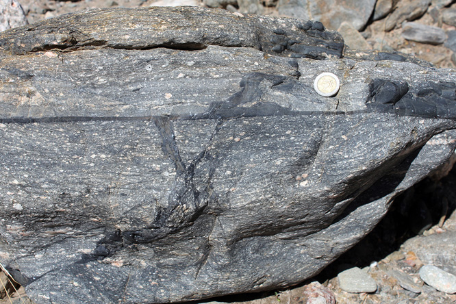
Pseudotachylites developed in ultramylonites of the Pichao-Ovejeria Shear Zone in Sierra de Quilmes, NW Argentina. From Monash University
.jpg)
mega-Pseudotachylyte. Vredefort impact crater, S. Africa. From Hartebeesthoek Radio Astronomy Observatory.
Bibliography
• David Shelley (1983): Igneous and metamorphic rocks under the microscope. Campman & Hall editori.
• E. WM. Heinrich (1956): Microscopic Petrografy. Mcgraw-hill book company,inc
• Anthony R. Phillpotts & Jay J. Ague: Principles of igneous and metamorphic petrology. Cambridge editore.
• Passchier, Cees W., Trouw, Rudolph A. J: Microtectonics (2005)
.jpg)
Pseudotachylyte (black) matrix in a deformed quartz-schist. PPL image, 2x (Field of view = 7mm) |
.jpg)
Pseudotachylyte (black) matrix in a deformed quartz-schist. PPL image, 2x (Field of view = 7mm) |
.jpg)
Pseudotachylyte (black) matrix in a deformed quartz-schist. PPL image, 2x (Field of view = 7mm) |
.jpg)
Pseudotachylyte (black) matrix in a deformed quartz-schist. PPL image, 2x (Field of view = 7mm) |
.jpg)
Pseudotachylyte (black) matrix in a deformed quartz-schist. PPL image, 2x (Field of view = 7mm) |
.jpg)
Pseudotachylyte (black) matrix in a deformed quartz-schist. PPL image, 2x (Field of view = 7mm) |
.jpg)
Pseudotachylyte (black) matrix in a deformed quartz-schist. PPL image, 2x (Field of view = 7mm) |
.jpg)
Pseudotachylyte (black) matrix in a deformed quartz-schist. PPL image, 2x (Field of view = 7mm) |
.jpg)
Pseudotachylyte (black) matrix in a deformed quartz-schist. PPL image, 2x (Field of view = 7mm) |
.jpg)
Pseudotachylyte (black) matrix in a deformed quartz-schist. PPL image, 2x (Field of view = 7mm) |
.jpg)
Pseudotachylyte (black) matrix in a deformed quartz-schist. PPL image, 2x (Field of view = 7mm) |
.jpg)
Pseudotachylyte (black) matrix in a deformed quartz-schist. PPL image, 2x (Field of view = 7mm) |
.jpg)
Pseudotachylyte (black) matrix in a deformed quartz-schist. PPL image, 2x (Field of view = 7mm) |
.jpg)
Pseudotachylyte (black) matrix in a deformed quartz-schist. PPL image, 2x (Field of view = 7mm) |
.jpg)
Pseudotachylyte (black) matrix in a deformed quartz-schist. PPL image, 2x (Field of view = 7mm) |
.jpg)
Pseudotachylyte (black) matrix in a deformed quartz-schist. PPL image, 2x (Field of view = 7mm) |
.jpg)
Pseudotachylyte (black) matrix in a deformed quartz-schist. PPL image, 2x (Field of view = 7mm) |