

Serpentine - Mg3Si2O5(OH)4
Serpentine was named in 1564 by Georgius Agrigola (Georg Bauer) from the Latin "serpens" = snake in allusion to the mottled green appearance of the mineral suggesting the resemblance to some snakes. The Serpentine group is composed of several related minerals. A generic formula that includes all members is:X2-3Si2O5(OH)4
Where X = Mg, Fe2+, Fe3+, Ni , Al, Zn, or Mn. One of the two Si atoms may also be replaced by an Al or Fe atom in a few rare members.
This leads to a complete formula of:
(Mg,Fe,Ni,Al,Zn,Mn)2-3(Si,Al,Fe)2O5(OH)4
The most common members of the serpentine group are:
Antigorite - (Mg,Fe)3Si2O5(OH)2
Chrysotile - Mg3Si2O5(OH)4
Lizardite - Mg3(Si2O5)(OH)4
Their differences are minor and almost indistinguishable in hand samples. However, the chrysotile minerals are more likely to form serpentine asbestos, while antigorite and lizardite form cryptocrystalline masses sometimes with a lamellar or micaceous character. All serpentine varieties have basically the same structure, a repeated two-layer arrangement of one tetrahedral (SiO4) and one octahedral (Mg(OH)2) layer. Three varieties of serpentine (Antigorite, Lizardite and chrysolite) are distinguished by physical deformation, disposition of the basic two-layer.
Serpentine minerals are always secondary, they occur together as alteration products of Al-poor, magnesian minerals, especially olivine, Mg-Pyroxenes, Mg-amphiboles. The most characteristic occurrence of serpentine is that derived from dunites, peridotites and other ultramafic-mafic rocks. A second occurrence of serpentine is in metamorphic contact zone in carbonate rocks, where it results from the alteration of forsterite marble.
Optical properties
• Form: Asbestiform, as parallel fibers ore aggregate with cross fibers.
• Color: Colorless to pale green
• Relief: Low.
• Interference colors: Low, grey, yellow.
Bibliography
• Bucher, K., & Grapes, R. (2011). Petrogenesis of metamorphic rocks. Springer Science & Business Media.
• Fossen, H. (2016). Structural geology. Cambridge University Press.
• Howie, R. A., Zussman, J., & Deer, W. (1992). An introduction to the rock-forming minerals (p. 696). Longman.
• Passchier, Cees W., Trouw, Rudolph A. J: Microtectonics (2005).
• Philpotts, A., & Ague, J. (2009). Principles of igneous and metamorphic petrology. Cambridge University Press.
• Shelley, D. (1993). Igneous and metamorphic rocks under the microscope: classification, textures, microstructures and mineral preferred-orientations.
• Vernon, R. H. & Clarke, G. L. (2008): Principles of Metamorphic Petrology. Cambridge University Press.
• Vernon, R. H. (2018). A practical guide to rock microstructure. Cambridge university press.
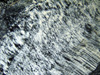
.jpg) Serpentine in a serpentinite. Todtmoos baden, Germany. XPL image, 10x (Field of view = 2mm) |
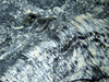
.jpg) Serpentine and talc in a serpentinite. Todtmoos baden, Germany. XPL image, 10x (Field of view = 2mm) |
.jpg) Serpentine in a serpentinite. Todtmoos baden, Germany. XPL image, 10x (Field of view = 2mm) |
.jpg) Serpentine in a serpentinite. Todtmoos baden, Germany. XPL image, 10x (Field of view = 2mm) |
.jpg) Serpentine and talc in a serpentinite. Todtmoos baden, Germany. XPL image, 10x (Field of view = 2mm) |
.jpg) Serpentine in a serpentinite. Todtmoos baden, Germany. XPL image, 10x (Field of view = 2mm) |
.jpg) Serpentine in a serpentinite. XPL image, 2x (Field of view = 7mm) |
.jpg) Serpentine in a serpentinite. XPL image, 10x (Field of view = 2mm) |
.jpg) Serpentine in a serpentinite. XPL image, 10x (Field of view = 2mm) |
.jpg) Serpentine in a serpentinite. XPL image, 10x (Field of view = 2mm) |
.jpg) Serpentine in a serpentinite. XPL image, 10x (Field of view = 2mm) |
.jpg) Serpentine in a serpentinite. XPL image, 40x (Field of view = 0.2mm) |
.jpg) Serpentine in a serpentinite. XPL image, 40x (Field of view = 0.2mm) |
 Serpentine in a serpentinite. XPL image, 10x (Field of view = 2mm) |
 Serpentine in a serpentinite. XPL image, 10x (Field of view = 2mm) |
.jpg) Mesh structure in olivine (high relief). The veins are filled by serpentine. PPL image, 10x (Field of view = 2mm) |
.jpg) Mesh structure in olivine (high relief). The veins are filled by serpentine. PPL image, 10x (Field of view = 2mm) |
.jpg) Mesh structure in olivine (high relief). The veins are filled by serpentine. XPL image, 10x (Field of view = 2mm) |
.jpg) Serpentine vein (grey) in a serpentinized peridotite. XPL image, 2x (Field of view = 7mm) |
.jpg) Serpentine vein (grey) in a serpentinized peridotite. XPL image, 10x (Field of view = 2mm) |
.jpg) Serpentine vein (grey) in a serpentinized peridotite. XPL image, 10x (Field of view = 2mm) |
.jpg) Serpentine vein (grey) in a serpentinized peridotite. XPL image, 10x (Field of view = 2mm) |
.jpg) Serpentine vein (grey) in a serpentinized peridotite. XPL image, 10x (Field of view = 2mm) |
.jpg) Serpentine vein (grey). XPL image, 40x (Field of view = 0.2mm) |
.jpg) Mesh structure in olivine. The veins are filled by serpentine and magnetite. PPL image, 2x (Field of view = 7mm) |
.jpg) Mesh structure in olivine. The veins are filled by serpentine and magnetite. PPL image, 2x (Field of view = 7mm) |
.jpg) Mesh structure in olivine. The veins are filled by serpentine and magnetite. XPL image, 2x (Field of view = 7mm) |
.jpg) Mesh structure in olivine. The veins are filled by serpentine and magnetite. PPL image, 2x (Field of view = 7mm) |
.jpg) Mesh structure in olivine. The veins are filled by serpentine and magnetite. XPL image, 2x (Field of view = 7mm) |
.jpg) Mesh structure in olivine. The veins are filled by serpentine and magnetite. PPL image, 2x (Field of view = 7mm) |
.jpg) Serpentine in a serpentinite. Todtmoos baden, Germany. XPL image, 10x (Field of view = 2mm) |
.jpg) Serpentine in a serpentinite. Todtmoos baden, Germany. XPL image, 10x (Field of view = 2mm) |