

Tourmaline - (Na,Ca)(Mg,Fe2+,Fe3+,Ti4+, Cr, Al, Li+)3Al6(BO3)3[Si6O18](OH)4
The name "tourmali" was a generic name used in Ceylon (Sri Lanka) for colored gems, mostly zircons. About 1703, it had been discovered by Dutch lapidaries that some of the "zircons" arriving in the Netherlands were actually a previously undescribed mineral. Several names were given to the new mineral including "Pierre de Ceylan, by Lemery in 1717. Tourmalin, as a more or less specific mineral name, was used by Rinmann in 1766.The structure of tourmaline consists of six-fold rings of silicon tetrahedra stacked with BO3 groups between the rings that form columns parallel to the c axis. Na ions reside in the center of the rings and Mg and other related elements are located along the inside edge of the rings. Al ions laterally tie together the stacks of rings in a distorted octahedral coordination. Ions in this complex silicate can undergo the following substitutions: Ca for Na in the ring centers; Mg and Al for Li in six-fold coordination between Si rings and BO3 groups; Fe3+ and Mn3+ for the Al links between Si rings.
The general formula is:
A(D)3G6(T6O18)(BO3)3Y3Z
Where
A = Ca, Na, K, or is vacant (large cations)
D = Al, Fe2+, Fe3+, Li, Mg2+, Mn2+ (intermediate to small cations - in valence balancing combinations when the A site is vacant);
G = Al, Cr3+, Fe3+, V3+ (small cations);
T = Si (and sometimes minor Al, B3+);
Y = O and/or OH;
Z = F, O and/or OH.
Boron is known or inferred to be an important component in many peraluminous silicic magmas that form granites, pegmatites, and rhyolites. Primary disseminated tourmaline may indicate the presence of boron in magma, but boron mineralization is commonly more abundant along the margins of intrusive rocks. Tourmaline may form in late-stage granites, pegmatites or during hydrothermal pneumatolitic introduction of boron. In granites tourmaline is usually schorl type, and it may form by late-stage alteration of granitic minerals by pneumatolitic fluid containing boron, and extreme tourmalinization may yield a rock of only tourmaline and quarz, like Luxullianite from the village of Luxulyan in Cornwall, England.
Optical Properties:
• Form: Tourmaline may be euhedral ranging from stubby columnar crystals to acicular crystals.
• Color: Highly variable and often irregular, brown, yellow, red, blue, green.
• Relief: Medium.
• Interference colors: III orders.
Tourmaline is distinguished from biotite and hornblende by the absence of cleavage, the presence of striated prisms, and (for hornblende) parallel extinction. Lighter colored tourmalines can be confused with topaz, apatite or corundum, but can be distinguished by certain optical properties. Topaz is biaxial, apatite has lower birefringence, and corundum has higher indices of refraction.
Minerals with trigonal symmetry, like tourmaline, show two specific absorption colours (dichroism), parallel to the vibration directions of the E-and O-waves. Sections orthogonal to the crystallographic c-axis (= optic axis) generally show the absorption colour of the O-wave as the microscope stage is rotated. Sections parallel to the c-axis show an alternation between the absorption colour of the E-wave (E-W orientation of the c-axis) and the O-wave (N-S orientation of the c-axis) for every 90°rotation of the stage.
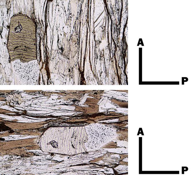
Change of absorption colour of tourmaline during the rotation of the microscope stage. A = Analyzer; P = Polarizer.
Bibliography
• Bucher, K., & Grapes, R. (2011). Petrogenesis of metamorphic rocks. Springer Science & Business Media.
• Fossen, H. (2016). Structural geology. Cambridge University Press.
• Howie, R. A., Zussman, J., & Deer, W. (1992). An introduction to the rock-forming minerals (p. 696). Longman.
• Passchier, Cees W., Trouw, Rudolph A. J: Microtectonics (2005).
• Philpotts, A., & Ague, J. (2009). Principles of igneous and metamorphic petrology. Cambridge University Press.
• Shelley, D. (1993). Igneous and metamorphic rocks under the microscope: classification, textures, microstructures and mineral preferred-orientations.
• Vernon, R. H. & Clarke, G. L. (2008): Principles of Metamorphic Petrology. Cambridge University Press.
• Vernon, R. H. (2018). A practical guide to rock microstructure. Cambridge university press.
.jpg) Green tourmaline and yellow staurolite crystals. Posada Asinara line, Sardinia (Italy). PPL image, 10x (Field of view = 2mm) |
.jpg) Basal section of green tourmaline. Posada Asinara line, Sardinia (Italy). PPL image, 10x (Field of view = 2mm) |
.jpg) Green tourmaline and yellow staurolite crystals. Posada Asinara line, Sardinia (Italy). PPL image, 10x (Field of view = 2mm) |
.jpg) Green tourmaline and yellow staurolite crystals. Posada Asinara line, Sardinia (Italy). PPL image, 10x (Field of view = 2mm) |
.jpg) Green tourmaline and yellow staurolite crystals. Posada Asinara line, Sardinia (Italy). PPL image, 10x (Field of view = 2mm) |
.jpg) Basal section of green tourmaline. Posada Asinara line, Sardinia (Italy). PPL image, 10x (Field of view = 2mm) |
.jpg) Basal section of green tourmaline. Posada Asinara line, Sardinia (Italy). PPL image, 10x (Field of view = 2mm) |
.jpg) Basal section of green tourmaline. Posada Asinara line, Sardinia (Italy). PPL image, 10x (Field of view = 2mm) |
.jpg) Fragmented torumaline crystal in a mylonite from Bhutan (Himalaya). PPL image, 2x (Field of view = 7mm) |
.jpg) Fragmented torumaline crystal in a mylonite from Bhutan (Himalaya). XPL image, 2x (Field of view = 7mm) |
.jpg) Fragmented torumaline crystal in a mylonite from Bhutan (Himalaya). XPL image, 2x (Field of view = 7mm) |
.jpg) Tourmaline crystals in a deformed granitic dyke. Capo Calamita promontory,Elba Island (Italy). PPL image, 2x (Field of view = 7mm) |
|
Tourmaline crystals in a deformed granitic dyke. Capo Calamita promontory,Elba Island (Italy). PPL image, 2x (Field of view = 7mm) |
.jpg) Tourmaline crystals (high interference colors) in a deformed granitic dyke. Capo Calamita promontory,Elba Island (Italy). XPL image, 2x (Field of view = 7mm) |
.jpg) Tourmaline crystals (in basal sections) in a deformed granitic dyke. Capo Calamita promontory,Elba Island (Italy). PPL image, 2x (Field of view = 7mm) |
.jpg) Tourmaline crystals in a deformed granitic dyke. Capo Calamita promontory,Elba Island (Italy). PPL image, 2x (Field of view = 7mm) |
.jpg) Tourmaline crystals (high interference colors) in a deformed granitic dyke. Capo Calamita promontory,Elba Island (Italy). XPL image, 2x (Field of view = 7mm) |
.jpg) Tourmaline crystals (basal and prismatic section) in a deformed granitic dyke. Capo Calamita promontory,Elba Island (Italy). PPL image, 2x (Field of view = 7mm) |
.jpg) Tourmaline crystals (basal and prismatic section) in a deformed granitic dyke. Capo Calamita promontory,Elba Island (Italy). XPL image, 2x (Field of view = 7mm) |
.jpg) Tourmaline crystals (basal section) in a deformed granitic dyke. Capo Calamita promontory,Elba Island (Italy). PPL image, 2x (Field of view = 7mm) |
.jpg) Tourmaline crystal (displaced by a cataclastic surface) in a deformed granitic dyke. Capo Calamita promontory,Elba Island (Italy). PPL image, 10x (Field of view = 2mm) |
.jpg) Tourmaline crystal (displaced by a cataclastic surface) in a deformed granitic dyke. Capo Calamita promontory,Elba Island (Italy). XPL image, 10x (Field of view = 2mm) |
.jpg) Broken Tourmaline crystal, in a deformed granitic dyke. Capo Calamita promontory,Elba Island (Italy). PPL image, 10x (Field of view = 2mm) |
.jpg) Broken Tourmaline crystal, in a deformed granitic dyke. Capo Calamita promontory,Elba Island (Italy). XPL image, 10x (Field of view = 2mm) |
.jpg) Zoned Tourmaline crystal, in a deformed granitic dyke. Capo Calamita promontory,Elba Island (Italy). PPL image, 10x (Field of view = 2mm) |
.jpg) Zoned Tourmaline in basal section, in a deformed granitic dyke. Capo Calamita promontory,Elba Island (Italy). PPL image, 10x (Field of view = 2mm) |
.jpg) Zoned Tourmaline in basal section, in a deformed granitic dyke. Capo Calamita promontory,Elba Island (Italy). PPL image, 10x (Field of view = 2mm) |
.jpg) Tourmaline crystals in a micaschist. PPL image, 10x (Field of view = 2mm) |
.jpg) Tourmaline crystals in a micaschist. XPL image, 10x (Field of view = 2mm) |
.jpg) Tourmaline crystals in a micaschist. PPL image, 10x (Field of view = 2mm) |
.jpg) Tourmaline crystals in a micaschist. XPL image, 10x (Field of view = 2mm) |
.jpg) Tourmaline crystals in a micaschist. PPL image, 10x (Field of view = 2mm) |
.jpg) Tourmaline crystals in a micaschist. XPL image, 10x (Field of view = 2mm) |
.jpg) Tourmaline crystals in a micaschist. PPL image, 10x (Field of view = 2mm) |
.jpg) Tourmaline crystals in a micaschist. XPL image, 10x (Field of view = 2mm) |
.jpg) Tourmaline crystals in a micaschist. PPL image, 10x (Field of view = 2mm) |
.jpg) Big tourmaline crystal (with internal foliation). PPL image, 1x (Field of view = 9mm) |
.jpg) Big tourmaline crystal (with internal foliation). XPL image, 1x (Field of view = 9mm) |
.jpg) Big tourmaline crystal (with internal foliation) and staurolite crystal. PPL image, 1x (Field of view = 9mm) |
.jpg) Big tourmaline crystal (with internal foliation) and staurolite crystal. XPL image, 1x (Field of view = 9mm) |
.jpg) Fragmented, big tourmaline crystal (with internal foliation). PPL image, 1x (Field of view = 9mm) |
.jpg) Fragmented, big tourmaline crystal (with internal foliation). XPL image, 1x (Field of view = 9mm) |
.jpg) Tourmaline crystal (with internal foliation). PPL image, 10x (Field of view = 2mm) |
.jpg) Tourmaline crystal (with internal foliation). XPL image, 10x (Field of view = 2mm) |