

Rutile - TiO2
Named in 1800 by Abraham Gottlob Werner from the Latin "rutilus", meaning "reddish", Rutile is a mineral composed primarily of titanium dioxide, TiO2. Rutile is the most common natural form of TiO2. Two rarer polymorphs of TiO2 are known:• Anatase (sometimes known by the obsolete name "octahedrite"), a tetragonal mineral of pseudo-octahedral habit.
• Brookite, an orthorhombic mineral.
The Rutile structure is the usual atomic arrangement for AX2 compounds with moderate-sixe cations. Each cation (Ti4+) is surrounded by six O2- ions in slightly deformed octahedral coordination and each O2- is bounded to three cations in triangular coordination. Rutile is basically TiO2 but forms some degree of solid solution with Tapiolite Fe(Nd,Ta)2O6. Fe2+, Fe3+, Ta5+ and Nb5+ may be present in rutule as major constituents. Rutile is more common than other TiO2 polymorph, it is dense, high-temperature, high-pressure mineral that occur in both igneous (granite, syenite, pegmatite) and metamorphic rocks (eclogite, marble).
Optical properties:
• Form: Usually more or less euhedral tetragonal crystals with square or octagonal section. It also occur as acicular hair-like inclusions in quartz or biotite.
• Color: Deep red-brown
• Relief: High
• Interference colors: Masked by mineral colors
Bibliography
• Bucher, K., & Grapes, R. (2011). Petrogenesis of metamorphic rocks. Springer Science & Business Media.
• Fossen, H. (2016). Structural geology. Cambridge University Press.
• Howie, R. A., Zussman, J., & Deer, W. (1992). An introduction to the rock-forming minerals (p. 696). Longman.
• Passchier, Cees W., Trouw, Rudolph A. J: Microtectonics (2005).
• Philpotts, A., & Ague, J. (2009). Principles of igneous and metamorphic petrology. Cambridge University Press.
• Shelley, D. (1993). Igneous and metamorphic rocks under the microscope: classification, textures, microstructures and mineral preferred-orientations.
• Vernon, R. H. & Clarke, G. L. (2008): Principles of Metamorphic Petrology. Cambridge University Press.
• Vernon, R. H. (2018). A practical guide to rock microstructure. Cambridge university press.
.jpg) Rutile (brown) crystals in an eclogite. PPL image, 10x (Field of view = 2mm) |
.jpg) Rutile (brown) crystals in an eclogite. PPL image, 10x (Field of view = 2mm) |
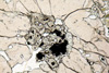
.jpg) Rutile crystals in an eclogite. XPL image, 10x (Field of view = 2mm) |
.jpg) Rutile (brown) crystals in an eclogite. PPL image, 10x (Field of view = 2mm) |
.jpg) Rutile crystals in an eclogite. XPL image, 10x (Field of view = 2mm) |
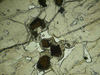
.jpg) Rutile (brown) with Titanite corona, enclosed in a Garnet. PPL image, 10x (Field of view = 2mm) |
 Rutile (brown) with Titanite corona, enclosed in a Garnet. PPL image, 10x (Field of view = 2mm) |
 Rutile (brown) with Titanite corona, enclosed in a Garnet. PPL image, 10x (Field of view = 2mm) |
.jpg) Rutile (brown) with Titanite corona, enclosed in a Garnet. PPL image, 10x (Field of view = 2mm) |
.jpg) Rutile (brown) with Titanite corona, enclosed in a Garnet. PPL image, 10x (Field of view = 2mm) |
(20).jpg) Rutile (brown) in a eclogite from Piedmont, Italy. PPL image, 10x (Field of view = 2mm) |
(13).jpg) Rutile (brown) in a eclogite from Piedmont, Italy. PPL image, 10x (Field of view = 2mm) |
.jpg) Rutile crystal (brown) in a kyanite schist. PPL image, 2x (Field of view = 7mm) |
.jpg) Rutile crystals (brown) and garnet and quartz crystals in a kyanite schist. PPL image, 2x (Field of view = 7mm) |
.jpg) Rutile crystals and garnet (isotropic) and quartz crystals in a kyanite schist. XPL image, 2x (Field of view = 7mm) |
.jpg) Rutile (brown) and kyanite crystal (right) in a kyanite schist. PPL image, 2x (Field of view = 7mm) |
.jpg) Rutile (brown) and kyanite crystal (right) in a kyanite schist. PPL image, 10x (Field of view = 2mm) |
.jpg) Rutile (brown) and kyanite crystal (to the top) in a kyanite schist. PPL image, 10x (Field of view = 2mm) |
.jpg) Rutile crystals (brown) to the edge of a granet in a kyanite schist. PPL image, 10x (Field of view = 2mm) |
.jpg) Rutile crystals to the edge of a granet (isotropic) in a kyanite schist. XPL image, 10x (Field of view = 2mm) |
.jpg) Rutile crystal (brown) to the edge of a granet in a kyanite schist. PPL image, 10x (Field of view = 2mm) |
.jpg) Rutile crystal (brown) to the edge of a kyanite crystal in a kyanite schist. PPL image, 10x (Field of view = 2mm) |
.jpg) Rutile crystal to the edge of a kyanite crystal (gray) in a kyanite schist. XPL image, 10x (Field of view = 2mm) |
.jpg) Rutile crystals (brown) enclosed in a garnet in a kyanite schist. PPL image, 20x (Field of view = 1mm) |
.jpg) Rutile crystal (brown) enclosed in a garnet in a kyanite schist. PPL image, 20x (Field of view = 1mm) |
.jpg) Rutile crystals (brown) in a kyanite schist. PPL image, 20x (Field of view = 1mm) |
.jpg) Rutile crystals (brown) in a kyanite schist. PPL image, 20x (Field of view = 1mm) |
.jpg) Rutile crystal (brown) in a kyanite schist. PPL image, 20x (Field of view = 1mm) |
.jpg) Rutile crystal (brown) in a kyanite schist. PPL image, 20x (Field of view = 1mm) |
.jpg) Rutile crystal (brown) in a kyanite schist. PPL image, 20x (Field of view = 1mm) |